Glossy Full Color Sandstone

Putative Tailspike Protein of a Bacteriophage (Vol
Made by
Print With Shapeways
 Choose Your Size
 Choose Your Material
 Choose your color and finish
$571.24
Have a question about this product?
contact the designerYou must be logged in and verified to contact the designer.
Product Description
Model Description
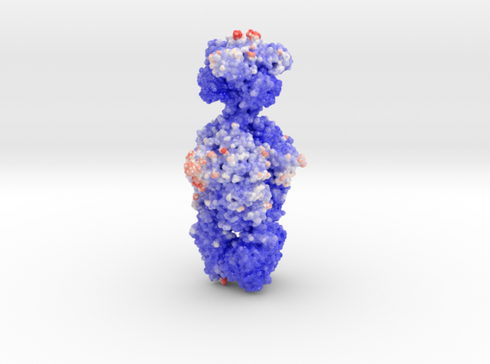
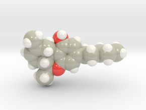
Here we see the volumetric surface of Putative Tailspike Protein, the lancing "weapon" of the virus. This model was created from PDB ID: 4oj5. The bulb at the top of the spear is the binding domain of the virus. Through the middle of the long sheath is a shaft that viral DNA flows through during viral infection.
The model is colored by the protein's thermic property (blue: cool, red: warm, white: neutral). As described in the pubmed description, the tailspike protein demonstrates strong thermic resistance which we can see in this molecular model.
Organism Description
Bacteriophages are viruses that infect and replicates within bacteria. This battle between virus and phage is an ancient war, waged from the beginning of life on our planet. Once these two foes locked into this battle for nuclear control, evolution produced a spectrum of amazing proteins like the well-known CRISPR systems for genetic reprogramming.
Protein Description
Bacteriophage tailspike proteins act as primary receptors, often possessing endoglycosidase activity toward bacterial lipopolysaccharides or other exopolysaccharides, which enable phage absorption and subsequent DNA injection into the host. Phage CBA120, a contractile long-tailed Viunalikevirus phage infects the virulent Escherichia coli O157:H7. This phage encodes four putative tailspike proteins exhibiting little amino acid sequence identity, whose biological roles and substrate specificities are unknown. Here we focus on the first tailspike, TSP1, encoded by the orf210 gene. We have discovered that TSP1 is resistant to protease degradation, exhibits high thermal stability, but does not cleave the O157 antigen. An immune-dot blot has shown that TSP1 binds strongly to non-O157:H7 E. coli cells and more weakly to K. pneumoniae cells, but exhibits little binding to E. coli O157:H7 strains. To facilitate structure-function studies, we have determined the crystal structure of TSP1 to a resolution limit of 1.8 Å. Similar to other tailspikes proteins, TSP1 assembles into elongated homotrimers. The receptor binding region of each subunit adopts a right-handed parallel ? helix, reminiscent yet not identical to several known tailspike structures. The structure of the N-terminal domain that binds to the virion particle has not been seen previously. Potential endoglycosidase catalytic sites at the three subunit interfaces contain two adjacent glutamic acids, unlike any catalytic machinery observed in other tailspikes. To identify potential sugar binding sites, the crystal structures of TSP1 in complexes with glucose, ?-maltose, or ?-lactose were determined. These structures revealed that each sugar binds in a different location and none of the environments appears consistent with an endoglycosidase catalytic site. Such sites may serve to bind sugar units of a yet to be identified bacterial exopolysaccharide.
https://www.rcsb.org/pdb/explore.do?structureId=4OJ5
Details
What's in the box:
Mdm: bacteriaphage 4oj5

Dimensions:
Success Rate:
First To try.
What's this?
Rating:
Mature audiences only.
